
Millions of tiny dots in vision – have you experienced this phenomenon? Scheerer’s Phenomenon, first described by Carl Scheerer in the late 19th century, is a natural and harmless occurrence caused by white blood cells moving through the capillaries in the retina. Most people experience it at some point in their lives, and in this article, we will explore its causes, characteristics, and potential as a diagnostic tool in ophthalmology. By the end of this article, you will have a better understanding of this unique visual phenomenon.

The difference between Floaters vs. tiny dots
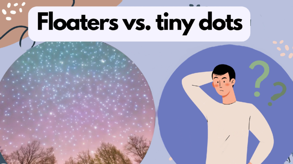
| Aspect | Floaters | Tiny Dots |
| Appearance | Gray or black specks, cobwebs, or strings | Pinpoint-sized dots or specks |
| Movement | Drift around, often following eye movements | May appear stationary or move slightly |
| Cause | Clumps of protein in vitreous humor | Dust particles, dirt, or debris |
| Commonness | Common in older individuals | Can occur in people of all ages |
| Seriousness | Usually harmless | Can be a sign of a retinal tear or detachment |
| Treatment | None required unless causing significant vision problems | None required unless caused by a medical condition |
| Prevention | None | Avoiding eye strain and protecting eyes from injury |
| Diagnostic test | Dilated eye exam | Visual examination or medical imaging tests |
The phenomenon known as Scheerer’s Phenomenon causes the appearance of tiny dots in vision.
Scheerer’s phenomenon occurs when light enters the eye and casts a shadow on the retina’s blood vessels. The blood vessels absorb the light, creating tiny dark spots that float across our vision. These spots are most visible against a bright background and appear as if they are moving in a swirling motion.
These dots are actually white blood cells moving through the blood vessels in the retina. The white blood cells, also known as leukocytes, travel through the capillaries that surround the retina. These capillaries are so small that the leukocytes need to squeeze through them, causing them to briefly block the light that enters the eye. The blocked light creates the tiny dark spots that we see.
The number of dots that we see can vary from person to person. It is estimated that on average, individuals can see between 5 and 10 dots per visual field. However, some people may see more, while others may not see any at all.
Scheerer’s phenomenon is a harmless and normal occurrence. In fact, it is a helpful tool for ophthalmologists to detect any abnormalities in the retina’s blood vessels. If there is a sudden increase in the number of dots seen, it may indicate a problem with the blood vessels, such as inflammation or blockage.
In conclusion, millions of tiny dots in vision, also known as Scheerer’s phenomenon, are white blood cells moving through the blood vessels in the retina. This phenomenon is harmless and normal, and while the number of dots varies from person to person, it is a helpful tool for detecting abnormalities in the retina’s blood vessels.
The millions of tiny dots in vision are visible due to the following reason.
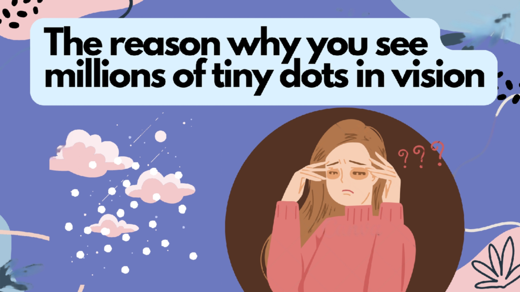
Here are some of the reasons why you see millions of tiny dots in vision:
- Aging: As people age, the vitreous gel inside the eye can shrink and become stringy, forming clumps or strands that cast shadows on the retina. These clumps and strands can appear as floaters.
- Eye injury: A direct hit to the eye or head trauma can cause floaters to appear.
- Eye diseases: Certain eye diseases, such as diabetic retinopathy, can cause the growth of abnormal blood vessels in the eye that can lead to floaters. Retinal detachment can also cause floaters to appear.
- Medications: Certain medications, such as corticosteroids, can cause floaters to appear.
- Eye surgery: People who have undergone eye surgery, such as cataract surgery or a vitrectomy, may experience floaters as a result of the procedure.
- Eye inflammation: Inflammation of the eye, such as uveitis, can cause floaters to appear.
- Genetics: Some people may be predisposed to developing floaters due to their genetics.
While floaters are usually harmless and do not require treatment, it is important to seek medical attention if you suddenly notice a large number of floaters, experience flashes of light, or notice a sudden decrease in vision. These symptoms may be a sign of a retinal tear or detachment, which requires prompt medical attention to prevent permanent vision loss.
Blue Sky and millions of dots in vision
Have you ever noticed the millions of tiny dots in your vision when you look up at the sky?
These dots are known as floaters, and they are actually tiny pieces of protein that are suspended in the vitreous humor, the gel-like substance that fills the eye. They cast a shadow on the retina, which is what creates the illusion of seeing these dots or specks. Floaters can appear in different shapes and sizes, including circles, lines, or webs.
While floaters are a normal part of the aging process, they can also be a sign of a more serious condition, such as a retinal tear or detachment. If you notice a sudden increase in floaters, especially accompanied by flashes of light or a loss of peripheral vision, it is important to seek medical attention immediately.
Interestingly, the appearance of floaters can be more noticeable when looking at a bright blue sky, as the contrast between the dark specks and the bright background makes them stand out more. However, they can also be visible against other backgrounds, such as a white wall or computer screen.
While floaters are generally harmless and do not require treatment, some people find them to be a nuisance and can even interfere with their vision. In rare cases, surgery may be an option to remove them, but this should only be considered in severe cases and after consulting with an eye specialist.
FAQs
Is blue entoptic phenomenon normal?
Yes, the blue entoptic phenomenon is normal and experienced by most people. It is caused by the visual perception of the white blood cells moving within the capillaries of the retina. These cells can appear as tiny, moving dots, lines, or squiggles that seem to be floating in the visual field. The blue color of this phenomenon is due to the absorption of shorter, blue wavelengths of light by the vitreous humor of the eye.
Why do I sometimes see tiny moving dots?
The perception of tiny moving dots or specks in the visual field is a common occurrence and is usually due to the presence of floaters in the eye. Floaters are small specks or clumps of cells that float around in the vitreous humor of the eye and cast shadows on the retina, creating the appearance of tiny dots or lines. These floaters can be more noticeable when looking at a bright background and tend to move as the eye moves. While floaters are generally harmless, sudden changes in their appearance or an increase in their number may be a sign of a more serious condition, and medical attention should be sought.
What causes Scheerer’s phenomenon?
Scheerer’s phenomenon, also known as the entoptic phenomenon, is caused by the perception of the structures within the eye itself, such as the vitreous humor, retina, and blood vessels. These structures can create visual effects such as floating dots, lines, or flashes of light that move as the eye moves. The phenomenon is most noticeable against a bright background such as a clear blue sky or a white wall.
To summarize
Scheerer’s Phenomenon is a natural and harmless visual phenomenon that causes millions of tiny dots to appear in an individual’s visual field. This phenomenon is caused by white blood cells moving through the capillaries in the retina and can be observed when looking at a clear blue sky, a blank white wall, or any bright uniform background. Although it may appear concerning, Scheerer’s Phenomenon is a normal part of human vision and is experienced by most people at some point in their lives. Furthermore, it can be a useful diagnostic tool in ophthalmology as it allows doctors to observe the flow of blood through the retinal capillaries and detect abnormalities in the retina that may affect vision.